
Early oncological diagnosis is an element affecting the course and prognosis of cancer. It consists of tests performed at various stages of the disease process. Oncological diagnostics can take many forms, and the performance of specific tests depends on many factors. Starting from screening tests, used in patients with risk influenza without clinical symptoms of the disease, to i.a. genetic tests, tumor markers and imaging techniques. Cancer diagnostics offers a variety of methods to diagnose cancer. It is reasonable to say that the sooner the cancer is diagnosed and its clinical advancement, the greater the chances of establishing a therapeutic procedure that will result in effective treatment.
Screening in oncology
Screening tests (so-called screening) in oncology are aimed at detecting early neoplastic changes in people who do not have clinical symptoms of cancer, but at the same time they may develop cancer, because - according to data obtained from epidemiological studies - the probability of occurrence of a given cancer in them is big. Screening can lead to a preliminary diagnosis of cancer, which must then be confirmed by appropriate additional tests. This allows for therapy with a high probability of cure. Screening tests cover the population of people with the highest probability of developing a specific cancer. Thanks to these tests, the number of deaths in the population covered by the screening is primarily reduced. Screening tests and early detection of the disease may also result in a decrease in the incidence of malignant neoplasms. According to recommendations, screening tests in Poland are recommended in the diagnosis of breast cancer (mammography), cervical cancer (cytological examination) and colorectal cancer (colonoscopy).

Genetic research
Genetic testing plays an increasingly important role in the diagnosis and prognosis of cancer. They may vary depending on the material collection and marking method. They are directed to people who do not report any symptoms, but whose family history indicates a genetic risk of cancer.
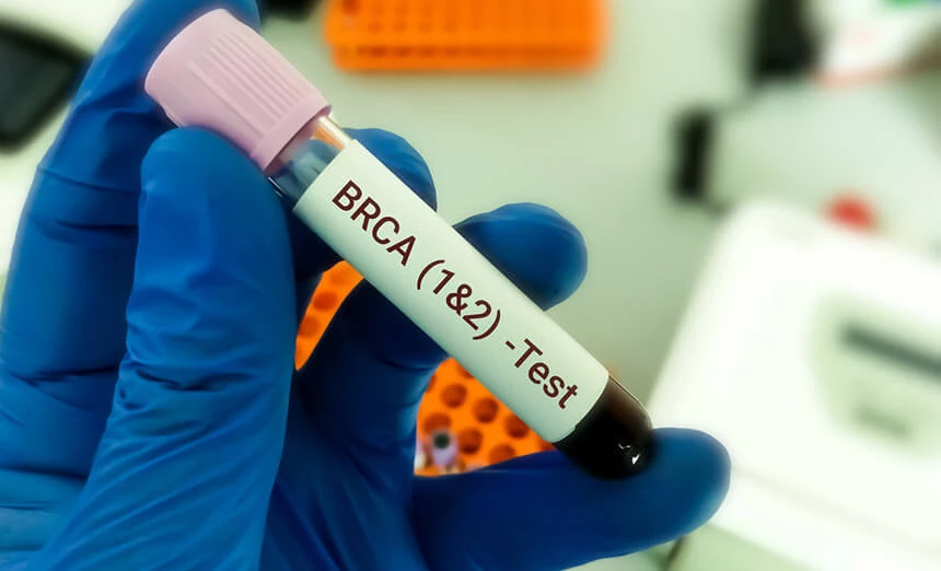
In this group of tests, it is worth paying attention to the BRCA1 and BRCA2 gene mutation testing. These genes are responsible for the repair mechanisms of damaged DNA. The appearance of a mutation on any of these genes indicates a failure to correct errors. As a consequence, the body is susceptible to the risk of cancer. The material for the test may be blood or a swab from the oral cavity.
Another test is liquid cytology and HPV DNA testing in the early detection of, among others: cervical cancer, the main cause of which is the HPV virus. Cytology with HPV genotyping is a test that allows to determine its oncogenicity and direct appropriate treatment.
Medical history and physical examination
A visit to a general practitioner gives a chance of diagnosing cancer at an early stage. Taking the history on the basis of the family history, lifestyle and symptoms reported by the patient (e.g. difficulty in swallowing, a palpable breast tumor or enlarged lymph nodes) prepares the ground for a physical examination. Depending on the medical history, the subject of the examination may include: skin, lymph nodes, head or abdominal cavity. Conclusions drawn on the basis of observations may be the basis for expanding diagnostic tests and an indication for the establishment of a DiLO green card.
Laboratory diagnosis of blood tests
Laboratory diagnostics plays a key role in oncology at every stage of cancer. It is used both when cancer is suspected and to monitor treatment. The most common tests include: haematological tests, determination of tumor markers, enzymes and a proteinogram.
Hematology tests may indicate active malignancy in patients who are anemic. At a later stage of the disease, they are performed to, for example, monitor the effects of cytostatic treatment.
Some cancers produce biological substances that can be quantified using tumor markers. Although they are not screening tests, they may suggest the presence of an inherited mutation that increases the risk of developing cancer. They are also of prognostic importance in initial diagnosis, therapy monitoring and post-treatment follow-up.
The determination of selected enzymes in the blood can help determine the stage of the cancer or indicate the presence of metastases.
Proteinogram is a test based on electrophoretic separation of blood proteins. In some cancers, some protein fractions are increased, and in advanced cancer, low albumin levels and hypoproteinization are often observed.
Liquid blood biopsy enables diagnosis of neoplastic disease, selection of therapy and monitoring of its effects in a sample of the patient's peripheral blood. This method detects circulating tumor cells, free circulating nucleic acids, exosomes and platelets produced by the tumor in a blood sample.

Picture diagnosis
One of the most commonly used diagnostic methods in oncology is diagnostic imaging. Due to the technical diversity of the medical equipment used and the method of obtaining the image, it can be divided into several types.
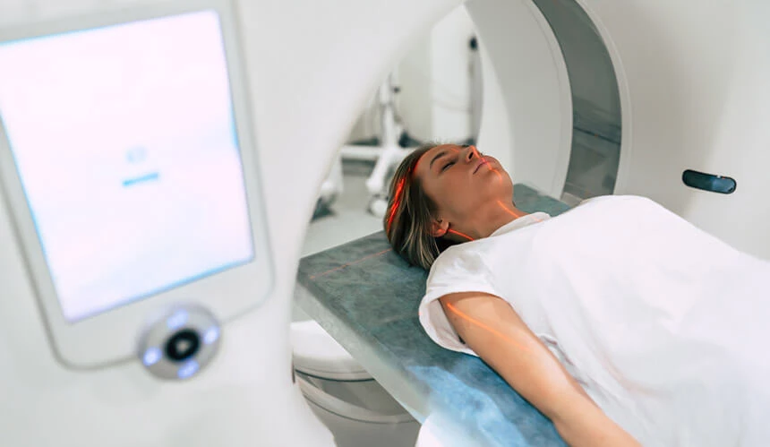
X-ray examinations, which include X-ray and computed tomography (CT), can be performed with the administration of a contrast medium, which in the case of computed tomography allows for, among others, blood vessel angiography. Mammography is a test that uses X-rays in the diagnosis of breast cancer. Computed tomography using a special spiral apparatus allows for the study of blood flow in the capillaries of a selected area (e.g. brain). Low-dose computed tomography (LDCT), thanks to a lower dose of radiation and more precise image quality, is a test used to diagnose early neoplastic changes in the lungs.
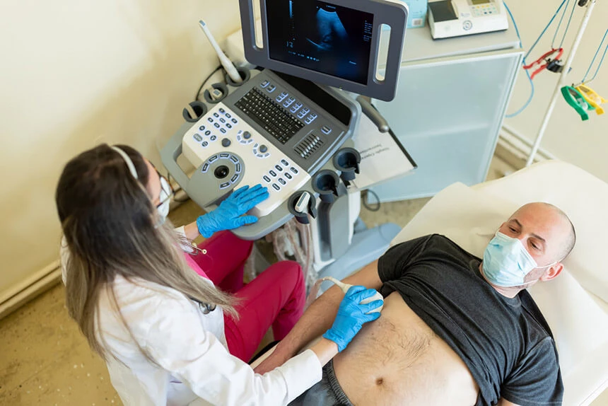
Imaging studies may also use ultrasonic waves, as in the case of USG, which allows to obtain a cross-sectional image of the examined object, or rely on the phenomenon of nuclear magnetic resonance (MR). In selected MRI studies, a contrast agent may be used to highlight the structures of individual organs. Magnetic resonance spectroscopy allows not only to visualize the examined organ (or tumor) in detail, but also to determine the degree of histological malignancy. This test also gives information about the biochemical composition in selected parts of the body - no need to do a biopsy. With both ultrasound and MR, arteries and veins can be examined (doppler ultrasound, MR perfusion).2
Advanced imaging diagnostics - radioisotope research and nuclear medicine
Isotopic labeling is used in oncological diagnostics to visualize the structures of organs imaged with gamma cameras.
Single photon emission tomography (SPECT) with the use of radiopharmaceuticals introduced into the body allows to obtain a three-dimensional image and precisely define metabolic foci.
Another test that makes certain organs visible is scintigraphy. It consists in graphic registration of the distribution of radioisotopes administered to the body. During their disintegration, they accumulate in the examined organ, which enables its precise morphological and functional assessment.
With positron emission tomography (PET), the radioactive substance given to the patient breaks down, producing radiation. As a result, photons are produced. They are recorded by detectors that enable the construction of images showing cross-sections of the patient's body, analogous to those obtained during magnetic resonance imaging.
Endoscopy
Endoscopic examinations find an important diagnostic application in the suspicion of neoplastic diseases. The role of endoscopy begins at the screening stage. It is also used to control the patient after oncological treatment, and in the case of early forms of cancer or precancerous conditions - in diagnostic and therapeutic procedures. Depending on the location of the body interior viewed, we distinguish specific types of endoscopy.
Gastroscopy involves an endoscopic examination of the upper digestive tract with the esophagus, stomach and duodenum. This makes it possible to visualize neoplastic changes on the membrane lining the lumen of these organs. An examination involving endoscopy of the lower gastrointestinal tract is called a colonoscopy. Endoscopic retrograde cholangiography (ERCP) allows the bile ducts and pancreatic duct to be contrasted with a duodenoscope. In the case of bronchoscopy, the patient has an examination of the upper airways using a bronchoscope inserted through the nose or mouth. Cystoscopy, on the other hand, allows for the assessment of the bladder mucosa after inserting a device called a cystoscope through the urethra. The equipment used in endoscopy can also be used to perform procedures (e.g. stopping bleeding, dilating strictures, removing a foreign body).
Histopathological diagnosis
Histopathological examination consists in microscopic examination of the collected cytological (cellular) material or histological material (tissue fragment). It allows for the detection and detailed diagnosis of cancer. The material for histopathological examination is most often collected during a biopsy or by means of exfoliative cytology. Histopathological examination includes, among others: completely excised lesions (cancer tumors), larger sections (taken during biopsy) or oligobiopsies, i.e. very small sections taken during endoscopy. In urgent mode, the so-called ad hoc (intraoperative) histopathological examinations. They are performed during the procedure to decide on its type (e.g. radical surgery). The choice of treatment method and prognosis depend on the result of the histopathological examination.
Summary
Type of examination | Name of study (or type of group of studies) | Examples of cancer diseases or selected applications in cancer diagnosis |
X-ray imaging studies | X-ray picture (X-ray) | Examination of various parts of the body, e.g. chest, spine. Bone cancer (metastasis), such as prostate cancer Lung cancer
|
Mammography | Breast cancer
| |
Computed tomography (CT) | The subject of examination is most often: brain, abdominal organs (liver, pancreas, spleen, vascular system, digestive tract), retroperitoneal space, skeletal system. Lung cancer Meningioma (brain cancer) Laryngeal cancer Salivary gland cancer Pleural mesothelioma Hepatocellular carcinoma Pancreatic cancer Lymphoma | |
Imaging studies without the use of X-rays | Ultrasound (ultrasound) transrectal, transvaginal, transesophageal, intravascular | Evaluation of the structure of the examined organ, as well as vascularity and flow within the tumor Thyroid cancer Salivary gland cancer Hemangioma of the liver Gallbladder cancer Pancreatic tumor Kidney tumor Tumor of the ovary, fallopian tubes |
Magnetic Resonance (RM) | Functional tests: - assessment of blood flow in the vessels - assessment of circulatory dynamics in tumors - assessment of the activity of the cerebral cortex in the vicinity of the neoplastic processGlejak mózgu Meningioma Vestibular-cochlear neuroma Pituitary adenoma Cervical body paraganglioma Intramedullary tumor | |
RM spectroscopy | Advanced diagnosis of tumors of the central nervous system (CNS) | |
Radioisotope research and nuclear medicine | Single photon emission computed tomography (SPECT) | Monitoring the course of the disease and assessing the effectiveness of treatment. Thyroid cancer, neuroendocrine tumors |
Scintigraphy | Bone cancer | |
Positron emission tomography (PET) | Searching for the primary focus of the disease and assessing the extent of the neoplastic process, determining the nature of the lesion: benign or malignant, detecting possible cancer metastases | |
Endoscopy | Gastroscopy | Stomach cancer |
Colonoscopy | Colon cancer | |
Endoscopic retrograde cholangiography (ERCP) | Pancreatic cancer, bile duct cancer | |
Bronchoscopy | Lung cancer | |
Cystoscopy | Bladder cancer | |
Blood laboratory tests | Hematology tests | Leukemia |
Tumor markers | Prostate cancer (PSA), ovarian cancer (CA 125) | |
Enzymes/Hormones | Testicular cancer (Beta HCG), Thyroid cancer (thyroglobulin, calcitonin) | |
Genetic research | the patient's DNA | Ovarian cancer (BRA1, BRCA2) |
oncogenic virus genetic material | Cervical cancer (HPV DNA test) | |
Biopsy | Fine Needle Aspiration Biopsy (FNAB) Fine-needle targeted aspiration biopsy (FNAB) Core biopsy Excisional and excisional biopsy | Collection of biological material from presumably diseased tissues, which is then evaluated in a histopathological examination. |
Histopathology |
| In the assessment of neoplastic diseases, in order to determine, among others, histological form, malignancy, tumor stage. |

