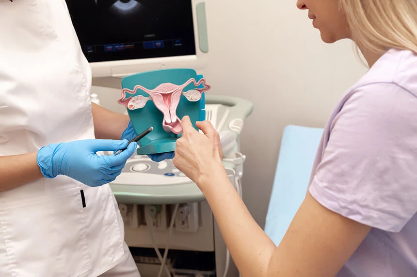
Two types of tumors can develop in the uterus, depending on where they originate:
- endometrial cancer, which most often develops in the mucous membrane lining the inside of the womb,
- cervical cancer
Endometrial cancer is most common in women between the ages of 55 and 70, but it occurs at a much younger age (5% of patients are under 40). It accounts for 7% of female cancers. The number of women suffering from the disease is increasing (in 2020, it was detected in almost 10,000 Polish women), but thanks to improved diagnostics and treatment, more of them are cured and the mortality rate does not increase.
Symptoms of uterine cancer
The typical symptoms of endometrial cancer include:
- bleeding from the genital tract outside menstruation - heavy and irregular
- bleeding after menopause
- blood-tinged discharge
- pelvic and sacral pain (appears in the advanced stage)
Less specific symptoms that can also indicate many other diseases include:
- weight loss
- weakness
The symptoms that most often prompt women to visit a doctor are bleeding and spotting in the postmenopausal period. Due to the fact that they cause anxiety, women usually seek medical attention quickly and cancer is detected at an early stage, when the prognosis is best.
Causes of endometrial cancer
The most common cause of the development of the disease is:
- excessive estrogen stimulation (both endogenous and exogenous), with simultaneous deficiency of progestogens
- excessive body weight
- a family history of endometrial cancer, colorectal cancer or breast cancer
- hormonal disorders caused by ovarian tumors
- postmenopausal age
- early menarche
- late age of last menstrual period
- no offspring
- occurrence of anovulatory cycles
- Polycystic ovary syndrome
- diabetes
- hypertension
- presence of congenital predisposition syndromes (Lynch syndrome, Cowden syndrome)
Diagnosis of endometrial cancer
In the event of disturbing symptoms, you should immediately go to the doctor. The basic element of diagnostics will be:
- gynecological examination
- endometrial abrasion (biopsy) with curettage of the cervical canal
- hysteroscopy
- an ultrasound examination of the reproductive organs and the abdominal cavity may be a possible supplement
- chest x-ray
- computed tomography or magnetic resonance imaging of the abdomen and pelvis
- basic blood and urine tests
Abnormalities in the uterine mucosa can be detected already in a transvaginal ultrasound examination. It allows the detection of even precancerous changes, i.e. those on which there is a high probability of cancer formation. This is a very important diagnostic method that can draw many women's attention to the fact that something is wrong in their body.
Endometrial abrasion involves histopathological examination of scrapings from the uterine cavity. The procedure is performed through the vagina under general anesthesia. After the procedure, the patient, if she feels well, goes home after a few hours.
Hysteroscopy, on the other hand, involves inserting a small camera into the uterine cavity, which allows you to view the endometrium at a very high magnification. Targeted samples are taken during the examination. This is an increasingly popular method.
Stage of endometrial cancer
The FIGO classification is used to determine the stage of advancement, in which stage I means the least advanced disease, and stage IV - the most advanced. Determining the stage of the disease is important in terms of treatment and prognosis. In order to determine the degree of involvement of the surrounding internal organs and lymph nodes and other organs as accurately as possible, the assessment is made after surgery.
Degree | Rating |
I | The tumor is strongly confined to the uterine wall |
IA | No infiltration or an infiltration <50% of the muscle depth |
IB | Infiltration equal to or greater than 50% of the depth of the site
|
II | Cervical stroma infiltrates, but cancer doesn't extend beyond the uterus
|
III | Local and/or regional infiltrates
|
IIIA | The infiltrates are in the serosa and/or appendages
|
IIIB | Vaginal and/or parametric metastases |
IIIC | Metastasis to periaortic and/or pelvic lymph nodes
|
IIIC1 | Involved pelvic lymph nodes
|
IIIC2 | Pelvic lymph node involvement with periaortic lymph node involvement
|
IV | Bladder and/or rectal infiltration and/or distant metastasis
|
IVA | Infiltrates on the bladder and/or rectal mucosa
|
IVB | Distant metastases, including to the inguinal and/or abdominal lymph nodes
|
Additionally, there are two types of endometrial cancer:
- type I, which occurs around menopause, develops on the basis of endometrial hyperplasia. It has to do with hormonal stimulation. It's going well.
- type II, is a glandular serous carcinoma. It is unrelated to hormonal stimulation and has a worse prognosis than type I.
Treatment of endometrial cancer
The type of treatment depends on the stage of advancement. Currently, treatment is mainly used for:
- a surgical method
- radiotherapy
- brachytherapy
- high-dose progesterone hormone therapy
- chemotherapy (in case of relapses)
Surgical treatment consists in removing the uterus along with the appendages. Alternatively, in justified cases, the surrounding lymph nodes are also removed. If the disease is very advanced, the operation is aimed at removing the cancerous tissue from the entire abdominal cavity as much as possible, the so-called maximum cytoreduction.
Surgical treatment is followed by adjuvant radiotherapy, which aims to destroy individual, imperceptible cancer cells. Such management increases the chances of treatment success and reduces the risk of vaginal metastases, but does not significantly affect 5-year survival.
In elderly women who suffer from circulatory diseases, surgery may be contraindicated. In this situation, brachytherapy is the treatment of choice. It consists in irradiating the lesions by placing the radiation source in the tumor or in its immediate vicinity (e.g. in the vagina). Unfortunately, studies show that brachytherapy alone gives worse results than surgical or combined treatment (surgery + radiotherapy).
Prognosis and complications of endometrial cancer
Among all gynecological malignancies, endometrial cancer has the best prognosis. 5-year survival rates are recorded:
- in 90% of women diagnosed with stage I cancer
- 60% in the second degree
- 40% in the third degree
- 5-9% in the 4th degree
Typical endometrial adenocarcinoma has a much better prognosis. This is the most common type of this cancer.
The most dangerous complication of endometrial cancer are metastases, which usually first appear in the lungs, liver and bones. Therefore, a chest X-ray is often ordered with this cancer to detect lung metastases. If disseminated cancer is suspected, then a CT scan or MRI should be performed.
Prevention of endometrial cancer
Prevention should primarily include pro-health behaviors. Reducing excessive body weight and taking care of the right amount of physical activity effectively reduce the risk of diabetes or hypertension (which are among the causes of cancer). In addition, it is believed that the right level of movement, which prevents obesity, reduces the risk of endometrial cancer by as much as half.
In addition, the risk of the disease is reduced in women using oral contraceptives or an intrauterine device, because they maintain hormonal balance.
Gynecological cancers and breast cancer account for more than half of cancer diagnoses in women. These are most often malignant tumors, and the possibility of curing depends on the stage of their advancement and early diagnosis. Sometimes careful self-observation is enough for a woman to pay attention to the first disturbing symptoms. Women should also remember about regular visits to the gynecologist and regular examinations - ultrasound, cytology. They help detect cancer at an early stage of development, when the chance of a full recovery is very high.
The presented medical information should not be treated as guidelines for medical conduct in relation to each patient. The medical procedure, including the scope and frequency of diagnostic tests and/or therapeutic procedures, is decided by the doctor individually, in accordance with medical indications, which he determines after getting acquainted with the patient's condition. The doctor makes the decision in consultation with the patient. If the patient wants to perform tests not covered by medical indications, the patient has the option of paying for them. |
Prezentowanych informacji o charakterze medycznym nie należy traktować jako wytycznych postępowania medycznego w stosunku do każdego pacjenta. O postępowaniu medycznym, w tym o zakresie i częstotliwości badań diagnostycznych i/lub procedur terapeutycznych decyduje lekarz indywidualnie, zgodnie ze wskazaniami medycznymi, które ustala po zapoznaniu się ze stanem pacjenta. Lekarz podejmuje decyzję w porozumieniu z pacjentem. W przypadku chęci realizacji badań nieobjętych wskazaniami lekarskimi, pacjent ma możliwość ich odpłatnego wykonania. Należy potwierdzić przy zakupie badania szczegóły do jego przygotowania. |
